
Case Intro
Whilst dawdling to finish the notes on a patient you’ve just seen, one of the nurses asks you to sign an ECG for a patient who’s just arrived.
ECG Interpretation
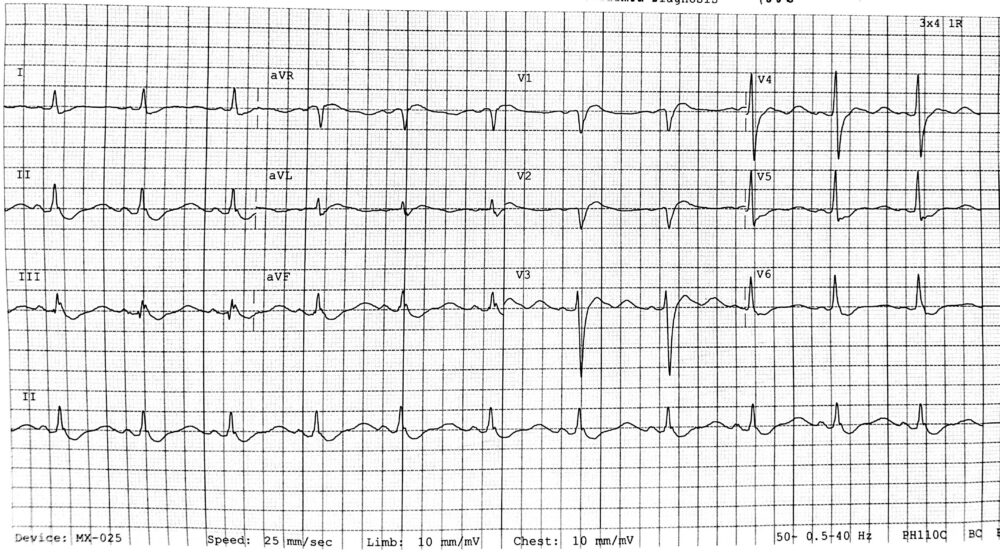
- Sinus rhythm
- Prolonged QRS suggestive of LBBB
- ST depression in the latero-inferior leads
- The paper QTc reads ‘705ms’ – this is a misread of the QT interval, caused by prominent U waves best seen in V3, reflective of QU prolongation.
U waves are classically caused by hypo-stuff; hypomagnesaemia, hypokalaemia, hypothermia (technically Osborne waves), as well as LVH and certain antiarrhythmics like digoxin, amiodarone, and sotalol. The first thing to check is a VBG for electrolyte derangement.
You go to talk to the 47 year old female patient. She has been feeling increasingly weak and tired over the past few weeks, as well as very thirsty and off her food. She drinks half a bottle of wine per day, and presented a few months ago with a similar picture. No vomiting, bowel changes, or medication changes.
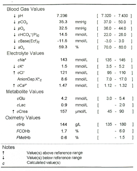
A VBG is pulled:
- Any acute life threats?
- What is the pH?
- What is driving this process?
- Is there adequate respiratory compensation?
- What is the anion gap?
- Any acute life threatening issues?
The potassium is only 1.5 mmol/l. Glucose is satisfactory at 4.2 mmol/l. - What is the pH?
7.23 indicates that there is an acidaemia. - What is driving this process?
The pCO2 is not raised, but the bicarbonate is low at 14.5 and the base excess is low at -11.5. This suggests a primarily metabolic acidosis. - Is there adequate respiratory compensation?
The low-normal pCO2 suggests some respiratory compensation.
Winters formula = (1.5 x 14.5) + 8 ±2 = 28-32 mmHg.
Given this is a venous blood gas, it is likely that an arterial gas would show adequate respiratory compensation. The patient’s work of breathing would be important to factor in. - What is the anion gap?
- Na – (Cl + HCO) = AG. Normal AG is 4-12 (when potassium not incorporated)
- 143 – (121 + 14.5) = 7.5
This is a normal anion gap acidosis (NAGMA).
The simplified strong ion difference (SID) calculation is another way to highlight a hyperchloraemic (NAGMA) driven acidosis.
Simplified SID = Na+ – Cl-
Simplified SID = 143-121 = 22 mmol/l
This is a substantially reduced SID from the typical range of 38-42, indicating that chloride is abnormally raised with a reduced bicarbonate due to the constraint of electroneutrality (as both are negatively charged ions)
Summary:
This is a pure normal anion gap metabolic acidosis with severe hypokalaemia.
Taking a look at each primary derangement individually before bringing it all together to diagnose the patient:
- Hypokalaemia
- Normal Anion Gap Metabolic Acidosis
Hypokalaemia
About 98% of total body potassium is intracellular, whilst the remaining 2% is in the extracellular fluid (reflected in serum measurements). This balance is maintained by the Na/K ATPase on cellular membranes, exchanging 3 Na+ out of the cell for 2 K+ into the cell, which helps establish the transmembrane potassium gradient for it to leak out and keep the cell membranes in a negative resting potential.
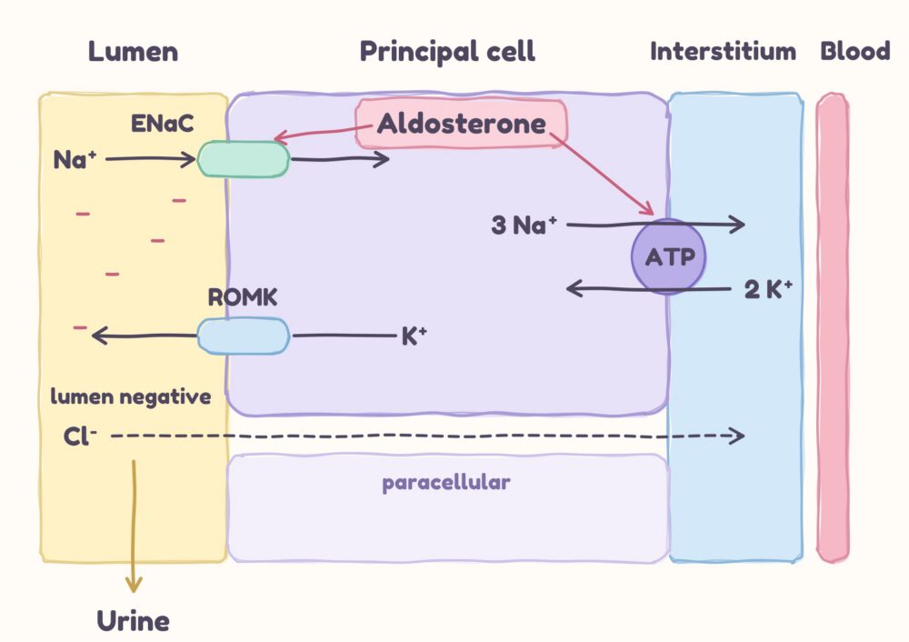
When potassium is filtered at the nephron, almost all of it is reabsorbed by the proximal tubules and loop of Henle, whilst the final urinary excretion of potassium is mediated by aldosterone at the principal cells in the distal tubules. Here, aldosterone stimulates Na+ absorption via epithelial sodium channels (ENaC), leading to K+ efflux through ROMK channels into the tubules, driven by the electrochemical gradient. Na+/K+-ATPase on the other side of the principal cells maintain the intracellular supply of K+ for this efflux.
There are three primary ways to lose potassium:
- Shifting potassium into cells; insulin, beta agonists, alkalosis can drive this
- Gastrointestinal losses from vomiting and diarrhoea
- Renal loss
- Reduced renal absorption in the tubules
- Increased distal tubule sodium delivery; Na⁺ enters through ENaC, creating a lumen-negative electrical potential that favours K⁺ secretion into tubules
- Increased aldosterone action will increase sodium reabsorption and potassium excretion
- Low magnesium impairs renal potassium conservation
Due to the above, serum potassium changes may not reflect total body potassium. For example, serum hypokalaemia from increased shifting into cells from, for example, hyperventilation-induced alkalosis, will maintain high total body potassium. In the case of DKA, serum potassium may be normal, but the large solute and fluid losses in the urine caused by hyperglycaemic osmotic drag will result in low intracellular, and consequently total body, potassium.
How can hypokalaemia cause weakness?
A low serum potassium creates a larger electrochemical gradient between the intracellular and extracellular compartments. Potassium diffusion increases out of the cell, resulting in a more negative resting membrane potential and hyper-polarising the cell.
A greater stimulus is then required reach threshold potential and trigger depolarisation. Significant, prolonged hypokalaemia can lead to profound weakness and even paralysis…providing the heart doesn’t give out first.
Why is hypokalaemia a problem for the heart?
Potassium is required for repolarisation of the cardiac myocyte back to its resting membrane potential. One would think that the increased concentration gradient from hypokalaemia would make it easier for potassium to leave the cells and for the myocytes to repolarise, but the converse is true.
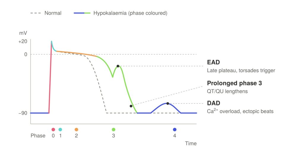
The key problems caused by low serum potassium are reduced potassium efflux (phase 3) and increased intracellular calcium (phase 2-3).
- The reduced extracellular K+ impairs several K+ channel currents used in repolarisation, leading to reduced K+ efflux from the cell.
- Reduced extracellular K+ results in reduced saturation of K+ binding sites of Na+/K+-ATPase, inhibiting Na+ extrusion and K+ uptake.
- This reduced transmembrane Na+ gradient impairs Na+/Ca2+ exchange, with less Na+ moving in and, therefore reduced Ca2+ extrusion out of the cell.
Prolonged action potential duration predisposes to early after depolarisations (EADs), which can trigger torsades de pointes, whilst intracellular Ca²⁺ overload and spontaneous sarcplasmic Ca²⁺ release predisposes to delayed after depolarisations (DADs), which can trigger ectopic activity.
Normal Anion Gap Metabolic Acidosis
Causes of normal anion gap acidosis include:
- Gastrointestinal tract (GIT) losses: Diarrhoea, high output stomas, GIT fistulas can cause large loss of electrolytes and bicarbonate.
- Iatrogenic chloride administration; such as sodium chloride
- Renal causes (Renal Tubular Acidosis)
- Impaired reabsorption of bicarbonate
- Impaired excretion of hydrogen ions
Once excluding GIT losses and excess sodium chloride administration, the remaining cause is likely to be some perturbation of renal function.
It’s worth recalling the basic anatomy & physiology of the kidneys to help determine what is going on. Regarding renal acid-base malfunction, the blame can usually be aportioned to either the proximal or distal nephron.
The nephron is the functional unit of the kidney. It is made up of:
- The renal corpuscle – filtration
- The renal tubules – reabsorption and secretion
- Proximal tubules – does most of the reabsorption
- Reabsorb ions (Na, K, PO, Ca, etc), glucose, amino acids, and organic acids.
- Reabsorb bicarbonate
- Produce ammonium
- Loop of Henle – Establishes medullary concentration gradient to concentrate urine
- Distal nephron – further regulates electrolyte, acid-base, water balance
- Distal tubules
- Reabsorb sodium and secrete potassium. Stimulated by aldosterone
- α-intercalated cells: hydrogen ion excretion
- Collecting ducts
- ADH/vasopressin and aquaporins → reabsorption of water
- Distal tubules
- Proximal tubules – does most of the reabsorption
In health, adults normally produce 15,000 mEq/day of volatile acids, and 1 mEq/kg/day of non-volatile acids from food These are reasonably arbitrary numbers, but nevertheless volatile acids are primarily excreted by the lungs, whereas the kidneys deal with the non-volatile acid load.
The kidneys regulate acid-base balance via:
- Regeneration & reabsorption of bicarbonate – proximal tubules
- The proximal tubules reabsorb around 80% of bicarbonate, whilst the rest is reabsorbed further in distal nephron, with almost none excreted in the urine.
- Excretion of hydrogen ions – distal nephron
- Type A intercalated cells in the distal nephron excrete hydrogen ions. Accumulating adequate free H+ in the urine would require extremely low urinary pH (<4.5). Instead, urinary buffers of ammonia (NH3) and phosphate (PO4-) bind excess H+ to circumvent this.
- Most phosphate (around 80%) is reabsorbed in the proximal tubules, with the remainder binding to secreted hydrogen ions in the distal nephron to be excreted. It is limited by how much is filtered, and has limited up regulation in acidosis. HPO₄²⁻ + H⁺ → H₂PO₄⁻
- Glutamine is an amino acid that is reabsorbed in the proximal tubules. It is metabolised in the proximal tubule epithelial cells to generate glutamate, ammonium (NH₄⁺) and bicarbonate (which is reabsorbed into the bloodstream). NH₄⁺ excreted in the proximal tubules is reabsorbed in the TAL as NH₃, which then diffuses back into the distal lumen before recombining with free hydrogen ions to form ammonium (NH₄⁺); from here it is trapped in the lumen and excreted in the urine. Ammonium is a buffer that can compensate; chronic acidosis can raise ammoniagenesis 5-10 fold. Phosphate can’t do this.
- Aldosterone: the action of aldosterone up-regulates ENaC activity and reabsorbs Na+ in the distal nephron. This increases negative charge within the distal lumen which favours H+ secretion into the tubules.
- Type A intercalated cells in the distal nephron excrete hydrogen ions. Accumulating adequate free H+ in the urine would require extremely low urinary pH (<4.5). Instead, urinary buffers of ammonia (NH3) and phosphate (PO4-) bind excess H+ to circumvent this.
So, renal tubular acidosis can be categorised into three distinct phenotypes:
- Distal nephron (RTA type I); dysfunction of hydrogen ion excretion
- Proximal nephron (RTA type II); dysfunction of bicarbonate reabsorption
- Aldosterone deficiency/resistance (RTA type IV)
Distal RTA (Type I)
- Location of the problem: Distal nephron; in the distal tubules and collecting ducts.
- Serum pH: In type I RTA, the α-intercalated cells cannot adequately excrete hydrogen ions into the tubular lumen, causing acidosis.
- Serum potassium: LOW. Hypokalaemia is due to renal K⁺ wasting. Altered distal nephron ion transport can promote K⁺ secretion; increased distal Na⁺ delivery and secondary hyperaldosteronism may contribute, particularly stimulated in the setting of volume depletion.
- Urinary pH: this is inappropriately raised (above pH 5.5) despite acidaemia, as the kidneys cannot adequately acidify the urine due to reduced H+ secretion and ammonium excretion.
- Urinary osmolar gap: this is usually low due to impaired ammonium excretion.
- Causes
- Autoimmune (sjogrens, SLE, RA, PBC, sarcoidosis, autoimmune thyroiditis)
- Nephrotoxins; lithium, trimethoprim, toluene
- Misc: Nephrocalcinosis (high Vit D, hyperparathyroidism), CKD, amyloidosis, intersitial nephritis
Proximal RTA (Type II)
- Location of the problem: Proximal tubules.
- Serum pH: In type II RTA, the proximal tubules have impaired bicarbonate reabsorption, causing an acidosis.
- Serum potassium: LOW. Coupled Na+ and HCO3- reabsorption is impaired in the proximal tubules, causing increased distal Na+, HCO3- and water delivery. The resulting urinary solute losses can contribute to volume depletion, activating the renin–angiotensin-aldosterone system. Aldosterone increases distal Na⁺ reabsorption and promotes K⁺ secretion. In addition to aldosterone action, increased Na⁺ delivery increases electrogenic Na⁺ reabsorption through ENaC makes the tubular lumen relatively negative, favouring K⁺ secretion down its electrochemical gradient.
- Urinary pH: this is reduced (generally below pH 5.5) as the kidneys can still acidify the urine by increasing H+ secretion
- Urinary osmolar gap: this is raised due to preserved/increased urinary NH₄⁺ excretion in response to the metabolic acidosis.
- Additional pearl: if proximal tubular dysfunction if more generalised than just bicarb reabsorption, it may lead to increased urinary loss of other solutes, protein, and glucose. This may show up as increased proteinuria, raised protein:creatinine / albumin:creatinine ratios, glycosuria, or hypophosphataemia, for example. Generalised proximal tubule reabsorption dysfunction is known as Fanconi Syndrome.
- Causes:
- Autoimmune; Sjogren
- Nephrotoxins:
- Carbonic anhydrase inhibitors: acetazolamide, topiramate
- Generalised defect: gentamicin, tetracycline, valproate
- Paraproteinaemia; multiple myeloma, amyloidosis, light chain deposits
- Genetic: Wilsons, Fanconi syndrome
Aldosterone resistance/deficiency (Type IV)
- Location of the problem: Distal nephron; in the distal tubules and collecting ducts
- Serum pH: Reduced. Hyperkalaemia suppresses renal ammoniagenesis, reducing NH₄⁺ excretion and net acid excretion. Reduced aldosterone activity also impairs distal acid excretion.
- Serum potassium: HIGH. Aldosterone deficiency or resistance leads to reduced ENaC activity and K+ secretion via ROMK channels. This causes retention of potassium and hyperkalaemia.
- Urinary pH: usually it is reduced (pH <5.5). Although ammonium generation is impaired, there is usually enough intact H+ ion secretion in the distal nephron to acidify the urine by excreting hydrogen ions.
- Urinary osmolar gap: this is usually low because ammonium excretion is impaired.
- Causes:
- Primary adrenal insufficiency; adrenal suppression (sepsis etc), autoimmune adrenalitis, congenital adrenal hyperplasia
- Meds:
- Renin-angiotensin-aldosterone blockade = ACEi, A2RB, potassium sparing diuretics
- NSAIDs
- Systemic disease; CKD, diabetes, adrenal insufficiency
| Feature | Distal (Type I) | Proximal (Type II) | Type IV |
| Defect | Impaired H+ excretion | Impaired HCO3- reabsorption | Aldosterone deficiency/resistance |
| Urinary pH | pH >5.5 | pH <5.5 | pH <5.5 |
| Serum potassium | Low-normal | Low-normal | Normal-high |
| Urinary osmolar gap | <150 mOsm | >150 mOsm | <150 mOsm |
| Urinary anion gap | Positive | Negative | Positive |
An Approach to Diagnosing RTA
The patient is presenting as a hyperchloraemic (normal anion gap) metabolic acidosis with profound hypokalaemia. We suspect this is renal in origin as she has had no GIT losses or new medications.
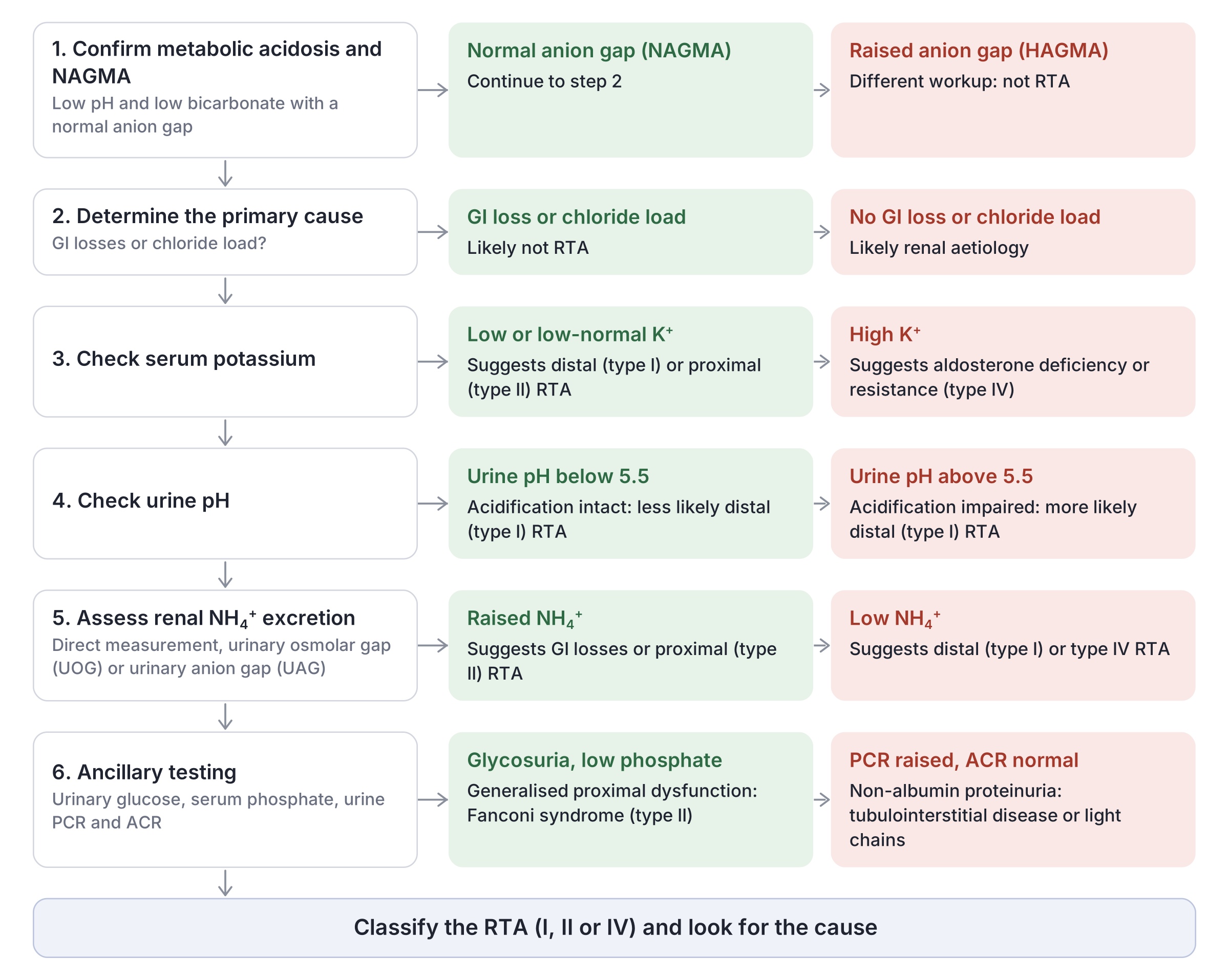
How do we determine what type of RTA is causing this? The following is a diagnostic approach:
1. Confirm a NAGMA
Use pH, anion gap, respiratory compensation calculation and, if helpful, delta ratio.
2. Determine the cause of the NAGMA
Gastrointestinal losses are most common; ask about vomiting, diarrhoea. If this hasn’t been a feature, and they haven’t been receiving other sources of chloride (eg saline), then this is likely a renal cause (ie RTA).
3. Check serum potassium
A LOW potassium will be congruent with a proximal (type II) or distal (type I) RTA. A HIGH potassium is in keeping with aldosterone deficiency or resistance (type IV RTA).
4. What is the urinary pH?
Low pH (<5.5) indicates that the kidneys are still able to acidify by excreting H+, which signifies the distal nephron apparatus is still working, thus suggesting a PROXIMAL RTA (Type II) or aldosterone deficiency/resistance (RTA type IV). On the other hand, inappropriately raised pH signifies that the distal nephron is unable to acidify the urine by excreting H+, which supports a DISTAL RTA (Type I)
5. Assess renal ammonium (NH₄⁺) excretion
The question to answer is: given this is an established metabolic acidosis, is the urinary NH₄⁺ excretion appropriately raised to compensate?
- A low urine ammonium level suggests that distal acidification is impaired, localising pathology to the distal nephron (type I), or due to impaired ammonium production from aldosterone deficiency (type IV) RTA.
- If the ammonium level is appropriately high, then this suggests proximal (type II) or GIT losses, as the distal nephron and aldosterone axis are likely still functioning ok to excrete ammonium (acid load).
There are three ways to measure urine ammonium:
- Direct urine NH₄⁺ measurement – may or may not be available
- Urinary osmolar gap (UOG)
- This is the difference between the measured and calculated osmolality.
- Calculated = 2 × (Na⁺ + K⁺) + urea + glucose
- Urine osmolality (measured) is the product of urinary K⁺, Na⁺, anions, urea, glucose and NH₄⁺ . Thus, the gap between the measured (which includes NH₄⁺) and the calculated (which does not include it) represents unmeasured urinary osmoses, of which ammonium salts may be a major contributor.
- UOG <150 mOsm/kg suggests diminished NH₄⁺ excretion due to DISTAL dysfunction (RTA type I) or ALDOSTERONE related (type IV) RTA. The lower the more certain.
- UOG >150 mOsm/kg suggests intact NH₄⁺ excretion, which points more toward a PROXIMAL (type II).
- Urinary anion gap (UAG)
- Urinary AG = (Na⁺ + K⁺) – Cl
- The question to ask; is the UAG positive or negative?
- The UAG is typically positive in health (between 20-90 mmol/L) when not needing to excrete large amounts of NH₄⁺, for example, to compensate for a metabolic acidosis.
- In the context of NAGMA:
- Positive UAG; signifies impaired NH₄⁺ excretion and suggests DISTAL (type I) or ALDOSTERONE-related (type IV) RTA.
- Negative UAG; suggests appropriately increased NH₄Cl excretion, which is a result of NH₄⁺ combining with chloride, to acidify the urine. This suggests PROXIMAL (type II).
6. Ancillary testing
Further testing can help determine whether this is more general impaired proximal tubule function, the findings of which may support a diagnosis of Fanconi syndrome. Remember that the proximal tubules reabsorb most of the solute, protein, and glucose that filters through the glomerulus.
- Urinary protein: a urinary protein:creatinine and albumin:creatinine ratio can help identify if there is increased protein loss in the urine. Albumin is one of the smallest measured proteins and so tends to appear first (though not always the case), whereas protein:creatinine ratio helps as a global marker of protein reabsorption dysfunction.
- Urinary glucose: glycosuria means that either the amount of filtered glucose at the glomerulus exceeds the reabsorption threshold (around 11.0 mmol/l), or that reabsorption is impaired due to proximal tubule dysfunction. Pair with serum glucose to discern general hyperglycaemia vs impaired reabsorption.
- Serum phosphate: a low serum phosphate, in the right clinical picture, may also indicate proximal tubular malabsorption.
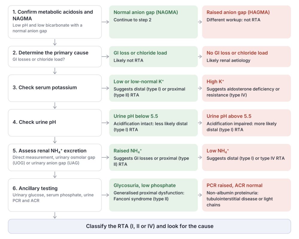
In the case of our patient:
- Confirm metabolic acidosis
- pH low, HCO low, pCO2 low-normal; confirms a metabolic acidosis
- AG 7.5 = NAGMA
- Determine primary cause – not GIT therefore more likely renal
- Check serum potassium
- Serum potassium is 1.5 mmol/l which supports RTA I or RTA II
- Incidentally, magnesium level was 1.16 mmol/L (high-normal)
- What is the urinary pH?
- The urinary pH was inappropriately high (pH 7.0), which indicates the kidneys are unable to acidify the urine by excreting H+ – this supports a DISTAL RTA (Type I)
- Assess renal ammonium (NH₄⁺) excretion
- Unable to perform direct measurement.
- UOG = urinary osmolality was not done(!)
- UAG
- = (Na⁺ + K⁺) – Cl
- = (31 + 19) – 27 = 23
- This is a marginally positive anion gap, which implies impaired renal ammonium excretion.
- Ancillary testing
- No glycosuria, normal phosphate level.
- Raised PCR, normal ACR. This suggests tubulointerstitial disease or light chains.
Bringing it all together:
This is a normal anion gap metabolic acidosis. There is severe hypokalaemia. The urinary pH is inappropriately raised. The anion gap is mildly positive, to suggest impaired ammonium excretion. The most likely differential is a distal (type 1) renal tubular acidosis.
At this stage the patient was already firmly out of the emergency department, with central potassium replacement using potassium acetate (to avoid extra chloride load). Screening for auto-immune disease and multiple myeloma were completed, as well as a renal ultrasound that showed some small non-obstructing stones.
Her hypokalaemia resolved over a few days, and inpatient renal biopsy was deferred in favour of outpatient workup. Sjogren syndrome remained a high differential after further enquiry revealed chronic sicca symptoms.
References
- Bhandari R, Ekladious A, Javaid MM. Demystifying normal-anion-gap metabolic acidosis: pathophysiology, aetiology, evaluation and diagnosis. Intern Med J. 2024;54:1056–65. doi:10.1111/imj.16418
- Bonner R, Hladik G. Renal tubular acidosis: core curriculum 2025. Am J Kidney Dis. 2025;85:501–12.
- Curthoys NP, Moe OW. Proximal tubule function and response to acidosis. Clin J Am Soc Nephrol. 2014;9(9):1627–38. doi:10.2215/CJN.10391012
- Das S, Agarwal V, Paul TV, Cherian KE. Approach to renal tubular acidosis – a review. Indian J Endocrinol Metab. 2026;30(1):26–36. doi:10.4103/ijem.ijem_774_25
- Ertek S, Caglar K. Underestimated reason of hyperkalemia in diabetic patients: type IV renal tubular acidosis – mini review. Front Clin Diabetes Healthc. 2025;6:1570868. doi:10.3389/fcdhc.2025.1570868
- Hoenig MP, Brooks CR, Hoorn EJ, Hall AM. Biology of the proximal tubule in body homeostasis and kidney disease. Nephrol Dial Transplant. 2025;40(2):234–43. doi:10.1093/ndt/gfae177
- Palmer BF, Kelepouris E, Clegg DJ. Renal tubular acidosis and management strategies: a narrative review. Adv Ther. 2021;38(2):949–68. doi:10.1007/s12325-020-01587-5
- Yaxley J, Pirrone C. Review of the diagnostic evaluation of renal tubular acidosis. Ochsner J. 2016;16(4):525–30. PMID: 27999512